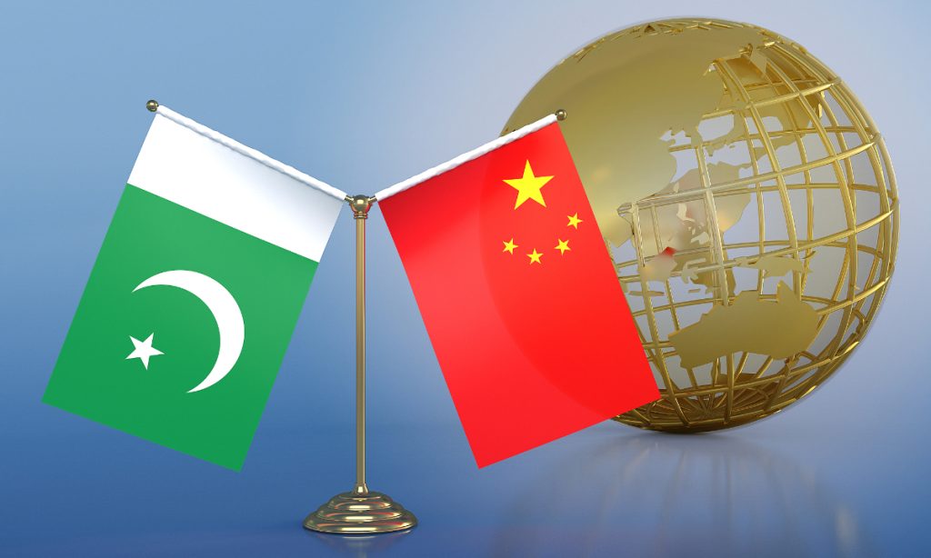
Chinese Premier Li Qiang on Monday kicked off his trip to Pakistan, where he is scheduled to attend the 23rd Meeting of the Council of Heads of Government of Member States of the Shanghai Cooperation Organization (SCO) to be held in Islamabad and pay an official visit to Pakistan from October 14 to 17, at the invitation of Prime Minister Muhammad Shehbaz Sharif of Pakistan.
Premier Li's visit to Pakistan is expected to strengthen and inject new momentum into the cooperation between Beijing and Islamabad, experts said.
"This is Premier Li's first visit to Pakistan after he took office and marks an exchange of visits at the head-of-government level between the two countries within a year, which is of significance to deepening all-weather strategic cooperative partnership," Mao Ning, spokesperson from China's Ministry of Foreign Affairs said on Monday.
Mao said China looks to working with Pakistan to enhance the traditional friendship, strengthen strategic communication, build the China-Pakistan Economic Corridor (CPEC) in a high quality way, deepen and expand cooperation across the board, ensure the safety and security of Chinese personnel, projects and institutions in Pakistan, accelerate the building of a closer China-Pakistan community with a shared future in the new era, and jointly safeguard regional peace, stability and prosperity.
Premier Li's visit is expected to further consolidate bilateral cooperation and push forward the next stage of development of CPEC, said Qian Feng, director of the research department at the National Strategy Institute at Tsinghua University.
However, the cooperation is currently being challenged by the threat of terrorism in Pakistan, which means that ensuring the safety of Chinese personnel and projects in Pakistan will likely be discussed during this visit, especially how to coordinate both China and Pakistan's efforts in fighting terrorism, said Cui Heng, a scholar from the Shanghai-based China National Institute for SCO International Exchange and Judicial Cooperation.
When asked for expectations of the 23rd Meeting of the Council of Heads of Government of Member States of the SCO, Mao said Premier Li will have an in-depth exchange of view with leaders of the participating countries on implementing consensus reached at the Astana summit and advancing SCO's practical cooperation.
Cui stated that the SCO meeting in Pakistan is expected to prioritize pragmatic cooperation in areas such as trade, investment, and people-to-people exchanges. "Security issues will also be on the agenda, as the region encompassing SCO member countries is facing increasing security threats while lacking a multilateral mechanism to address these challenges," Cui noted.
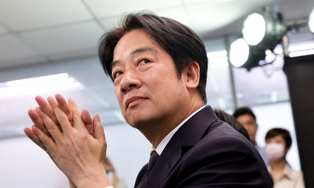
Taiwan regional leader Lai Ching-te's "Double Ten" speech is like "a poison pill wrapped in cellophane" and despite his repeated use of the word "peace," his propagation of the separatist "Taiwan independence" agenda remains unchanged, said analysts who noted that any actions toward secession will inevitably face decisive countermeasures.
On Sunday, the Center for Cross-Straits Relations Studies at the Renmin University of China held a seminar on changes in the political situation in Taiwan island and the direction of cross-Straits relations, during which scholars analyzed the evolution of Lai's separatist rhetoric since he assumed the role of Taiwan region's leader on May 20, leading up to his "Double Ten" speech. They said that despite the deceptive nature of his remarks, it is evident that Lai is not merely talking about "Taiwan independence" — he is actively taking steps to promote it.
Since Lai's speech on October 10, in which he claimed that the People's Republic of China has no right to represent Taiwan and that they are "not subordinate to each other," there has been widespread controversy and criticism from both the mainland people and people on the island. However, some Western media referred to Lai's "Double Ten" speech as "softer" than his recent speeches.
Wang Yingjin, director of the Center for Cross-Straits Relations Studies, said at the seminar on Sunday that Lai's "Double Ten" speech appeared to have introduced two changes: a noticeable reduction in provocative language and a shift in his stance toward the "Republic of China" (ROC) - moving from previously rejecting the "ROC" to now embracing and utilizing it.
The "ROC" Lai refers to, however, is one that has taken root specifically in the regions of Taiwan, Penghu, Kinmen, and Matsu - a "Taiwanized ROC." This is fundamentally distinct from the ROC advocated by the Kuomintang, which encompasses the mainland, said Wang, noting that these changes are merely tactical adjustments.
Lai's speech is highly deceptive and misleading, causing many in the international community to mistakenly perceive it as a "softening signal" toward the Chinese mainland, said Wang.
The core and essence of the cross-Straits portion of the "Double Ten" speech remained unchanged in six key aspects: the essence of promoting "Taiwan independence" has not changed; the confrontational stance toward the mainland has not changed; the narrative of playing the "democracy and freedom" card has not changed; the methods of colluding with external forces have not changed; the tactic of blaming the mainland for the deterioration of cross-Straits relations has not changed; and the hyping of the "China threat" rhetoric has not changed, Wang said.
"This all demonstrates that Lai is determined to recklessly advance along the path of 'Taiwan independence,' posing a serious threat to peace and stability in the Taiwan Straits. He is undoubtedly a 'pragmatic instigator of war,'" Wang told the Global Times.
Some scholars at the seminar described Lai's "Double Ten" speech, along with his recent separatist remarks that propagate a "two-state" rhetoric as "a poison pill wrapped in cellophane" or a "Taiwan independence stance under a new disguise." They emphasized that this cannot be seen as a so-called soft signal to the mainland but rather as a further move to sever cross-Strait relations that may led to more confrontations.
Lai's speech embodies the essence of "Taiwan separatism," laden with cunning and deceit. While some Western media depicted it as "soft," this supposed moderation is a mere façade. By emphasizing mutual non-subordination, Lai is attempting to legitimize "Taiwan separatism," said Zhu Guilan, an expert from the Institute of Taiwan Studies under Tsinghua University.
The current struggle against "Taiwan separatism" is not a matter of systems or ideologies, but a fight between reunification and separation, said Zhu Guilan, noting that "our fight is fundamentally about defending China's national sovereignty, which has never wavered over Taiwan island."
Lai and the Democratic Progressive Party's push for "Taiwan separatism" is entirely unjust, illegitimate and unlawful under both Chinese and international law, as well as among sovereign nations. His pushing of separatism, his efforts to seek foreign support - particularly from the US - and even his advocacy for military means to secure "Taiwan separatism" should face decisive countermeasures, Zhu Guilan said.
Although Lai frequently mentioned "peace" in his speech, his concept of peace is riddled with contradictions and hypocrisy. While he often invoked the term, his policies and rhetoric mask the true intent of fostering division and confrontation across the Taiwan Straits. His use of "peace" is insincere, serving instead as a tool to escalate tensions, Zhu Songling, a professor at the Institute of Taiwan Studies of Beijing Union University, told the Global Times on the sidelines of the Sunday seminar.
Zhu Songling noted that Lai and the separatists' actions in the island of Taiwan will inevitably challenge regional security and heighten the risk of war. Their "Taiwan independence" agenda will fail to protect Taiwan's security, putting its 23 million people in a precarious situation.
While the mainland is committed to peaceful reunification, it has made clear that any form of "Taiwan independence" will not be tolerated, said Zhu Songling, noting that timely warnings to Lai and his group are essential.
China’s Ministry of State Security (MSS) published an article on Friday to remind the public not to ignore the security of the idle network devices, as national security agencies discovered that overseas espionage organizations have been frequently targeting idle and discarded network equipment for cyberattacks in recent years, resulting in some network devices becoming “backdoors” for data leaks, posing a serious threat to China’s network security and data safety.
With the rapid development of the internet, the scale of network devices has grown exponentially. The swift evolution of network technology and applications has accelerated the iteration and upgrading of network equipment, leading to a significant rise in idle and discarded network devices, the MSS said in the article.
It was discovered that a decommissioned server belonging to a certain unit remained in the information technology room, creating an opportunity for overseas espionage agencies. These agencies gained control of the server through network scanning, subsequently infiltrating the internal network and using the decommissioned server as a springboard for cyberattack activities, according to the MSS.
A domestic camera monitoring platform was also found to have suffered an overseas cyberattack. Analysis revealed that the control platform’s server contained numerous usernames and passwords for the cameras it managed.
After being established, the platform remained idle, continuously powered on but without management, leading to high-risk issues such as long-term neglect, outdated system versions, and database vulnerabilities. If overseas espionage agencies could take control of this platform remotely, they could manipulate the relevant cameras for observation and potential intelligence theft, said the MSS.
In another case exposed by the MSS, a manufacturing company was found to have abnormal traffic transmission in a system developed by a third party. Upon investigation, it was found that the company had privately mapped multiple ports to the outside for easier system maintenance and had not closed them for a long time. Overseas espionage agencies scanned the mapped ports and used remote desktop access to log into the company’s system server, conducting cyberattacks, according to the MSS.
The MSS reminded the public to conduct comprehensive security precautions, thorough technical measures, and strengthen security education.
China's foreign exchange reserves totaled 3.3164 trillion U.S. dollars at the end of September, up by 28.2 billion dollars, or 0.86 percent compared to the end of August, official data showed Monday.
The State Administration of Foreign Exchange said that the U.S. dollar index declined, and global financial asset prices generally increased last month.
The combined effects of currency translation and asset price changes led to the increase in China's foreign exchange reserves in September, the administration added.
China has made significant progress in implementing the 102 key projects listed in its 14th Five-Year Plan (2021-2025), with notable achievements seen in sectors such as transportation infrastructure, energy systems and green development, the country's top economic planner has said.
According to the National Development and Reform Commission (NDRC), the following are select highlights of the achievements the projects have seen over recent years:
-- The country's transportation infrastructure network has been strengthened, with about 95 percent of the country's expressway services areas now equipped with charging facilities. A total of 151 national logistics hubs have been included on a list of key construction projects issued by the commission in 2021, covering 31 provincial-level regions.
-- Amid China's efforts to build a modern energy system, the world's largest clean energy corridor has been built along the Yangtze River. China's total installed power generation capacity has exceeded 3 billion kilowatts, with installed wind and solar power generation capacities surpassing coal power.
-- China has made coordinated progress in urban and rural development since 2021. In that time, it has begun renovating about 195,000 old urban residential communities, and approximately 210,000 kilometers of old gas pipelines have been renovated in urban areas.
The aggregate length of China's rural roads was 4.6 million kilometers by the end of 2023. Tap water coverage in China's rural areas had reached 90 percent by the end of last year, and about 75 percent of China's rural households had access to sanitary toilets.
-- The country has made systematic efforts to promote green development, with protected natural areas now accounting for about 18 percent of China's total land area. Led by China's broader efforts to cut pollution in key sectors, about 80 percent of the national steel production capacity has seen an ultra-low carbon emissions upgrade.
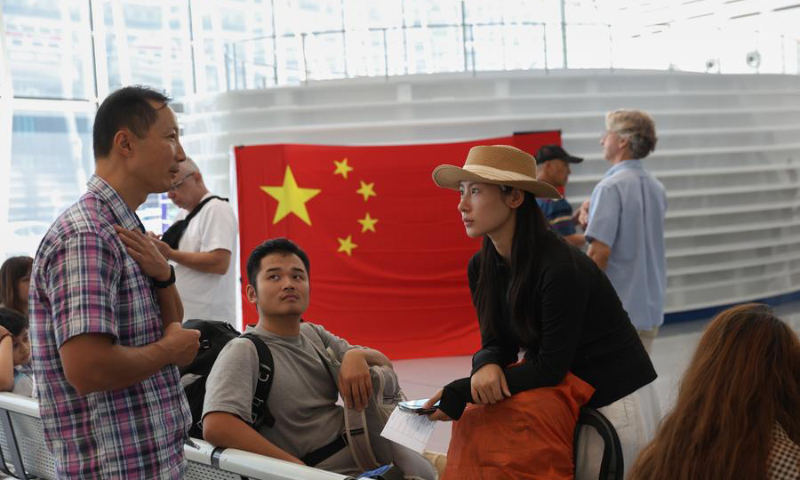
As the Lebanon-Israel conflict escalated, the Chinese government organized two batches of evacuation of overseas Chinese, which has aroused attention from all walks of life. According to a recent report of Jornal San Wa Ou, a Macao newspaper, during the evacuation, one of the evacuees was from China's Taiwan region. The report, citing Taiwan media, noted that even the "foreign affairs department" of the Taiwan island authorities admitted that with the help of the mainland, the Taiwan student left Lebanon aboard a cargo ship arranged by mainland authorities. The Taiwan authorities reportedly said it closely followed the process.
During the evacuation organized by China, Taiwan compatriots again received assistance, leading to political and media circles in Taiwan island debating the issue. Among them, Yuan Juzheng, a professor of philosophy in Taiwan, praised the mainland's diplomatic efforts during a program and suggested that if Taiwan residents "want to ensure their safety abroad, they should carry a Taiwan compatriot permit." He also said that every time there is a war abroad, the mainland's evacuation activities start very early. "I wonder how Taiwan people feel when they board mainland evacuation vehicles or ships and receive protection? The meaning of the evacuation explains everything."
Jornal San Wa Ou also highlighted instances where the mainland assisted Taiwan compatriots in evacuation operations. The report stated that for a long time, whenever the mainland initiated evacuation actions, they would help Taiwan compatriots who couldn't receive assistance from Taiwan authorities. The Chinese mainland has consistently treated Taiwan compatriots as Chinese citizens and evacuated them alongside mainland residents. For example, in June 2003, a civil war broke out in Liberia, prompting the Chinese mainland to initiate a evacuation operation, helping a total of 36 overseas Chinese, including Taiwan compatriots.
On April 18, 2006, riots occurred in the Solomon Islands, resulting in the burning of more than 60 Chinese shops in local Chinatown and severely impacting hundreds of overseas Chinese. The Chinese Foreign Ministry activated an emergency mechanism overnight, dispatching charter flights to evacuate these overseas Chinese in four batches from the Solomon Islands to Papua New Guinea, and then sending a government charter flight from the Chinese mainland to bring back 310 overseas Chinese to South China's Guangdong Province, among whom were also Taiwan compatriots.
In January 2008, fierce fighting broke out between anti-government forces and the Chadian government army, posing a serious threat to the lives and property of overseas Chinese and personnel of Chinese-funded institutions. The Chinese Foreign Ministry quickly activated an emergency mechanism, and with the full assistance of the Chinese embassy in Chad and Chinese embassies and consulates in France, Cameroon and Gabon, all 411 Chinese citizens were safely evacuated from Chad, including compatriots from Taiwan.
On September 4, 2018, after a typhoon and an earthquake struck Osaka, Japan, the Chinese Consulate General in Osaka assisted a total of 1,044 Chinese travelers stranded in Osaka, including 32 from Taiwan. In contrast, the Taiwan island's so-called representative office in Japan was criticized for its slow response, failing to provide timely assistance to Taiwan people. Many complained to the media that the office not only did not proactively assist during the incident but also stated that those in need had to email to request help. Some travelers reported that when they called the office, the staff had a poor attitude and did not provide assistance.
The report noted that the central government places great importance on ensuring that Taiwan compatriots are also assisted during the evacuation operations. Whether in the previous evacuation operation in Libya or in Ukraine, the Chinese leaders have issued guidance and directives. This spirit has also been incorporated into national policy documents. According to the 14th of the "26 measures" to benefit Taiwan, Taiwan compatriots can seek consular protection and assistance and apply for travel documents at embassies and consulates of the People's Republic of China overseas.
The report stated that the Chinese government's chartered evacuation flights not only reflect the concern and care of the Chinese leader and the central government for overseas Chinese but also demonstrate that China's national strength has developed to a point where it can provide safety and protection for overseas Chinese. The chartered evacuation indicates that China's people-first governance philosophy has been deeply integrated into the country's diplomatic policies. The inclusion of Taiwan compatriots in these operations underscores that the motherland is a strong support for all Chinese citizens, including those from Taiwan, the report added.
To celebrate the upcoming Mid-Autumn Festival, which will fall on Tuesday in 2024, the National Centre for the Performing Arts (NCPA) will present a series of exciting performances, including the Peking Opera Farewell My Concubine, which will be staged on Friday.
The opera is a classic work that was initially adapted from the story of Xiang Yu, the self-styled "Hegemon-King of Western Chu" from over 2,000 years ago, and his tragic farewell to his beloved concubine Yu Ji after being defeated. The story is based on a historical event from the late Qin Dynasty (221BC-206BC).
This opera was famously performed by Peking Opera master Mei Lanfang (1894-1961), who played the role of Yu Ji.
The opera has been performed by numerous prominent artists since its debut in 1918.
The performance structure was refined over time, eventually evolving into the eight-act version that audiences know today.
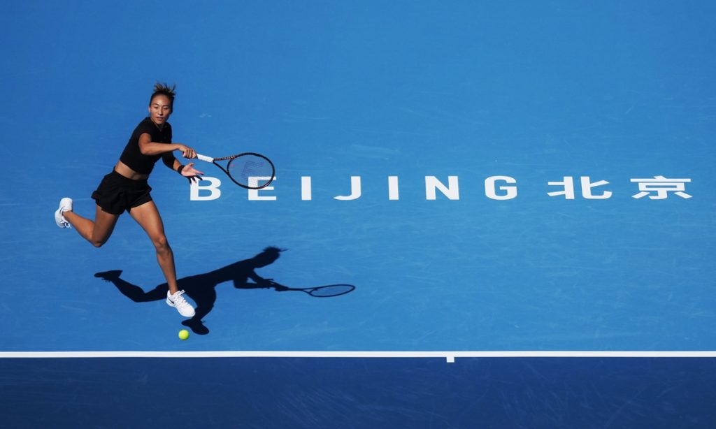
Chinese tennis fans are expecting the female Olympic gold medalist Zheng Qinwen to deliver quality performances at the upcoming China Open in Beijing, as the annual tennis event kicks off on Monday.
Since winning the Olympic women's singles gold medal, world No.7 Zheng has captured the spotlight in Chinese tennis.
Though the main draw has yet to be unveiled, Zheng, as a seeded player at the China Open, is possible to receive a bye in the first round and will play her opening match later this week.
Zheng's goal for the Chinese season is clear: to earn as many ranking points as possible to improve her chances of qualifying for the WTA Finals.
To secure one of the eight spots in the WTA Finals, Zheng will need to perform well at the China Open (WTA 1000), Wuhan Open (WTA 1000), and Ningbo Open (WTA 500), as she currently sits at No.9 in the race to the Finals, trailing Emma Navarro and Danielle Collins by 498 and 108 points respectively.
With Collins opting out of the China Open, Zheng has one less competitor in the fight for a coveted WTA Finals berth, a year after Zheng's disappointing first-round exit at the China Open last year.
Having returned to Beijing as early as Sunday to begin her preparations, Zheng's early training highlights her determination to perform well throughout the China swing. Strong performances during the China swing could secure her first WTA Finals appearance.
In the singles draw, world No.1 Iga Swiatek and world No.2 Aryna Sabalenka have already secured their places at the WTA Finals, leaving six spots still available. Swiatek will not participate in the China Open.
In the men's competition at the China Open, world top-three players Jannik Sinner of Italy, Alexander Zverev of Germany and Carlos Alcaraz of Spain are the marquee names of the list, with home favorites Zhang Zhizhen and Wu Yibing leading the Chinese squad's charge at the ATP 500 event.
This year's China Open singles qualifying rounds will take place on Monday and Tuesday, with the expanded 96-player singles main draw kicking off on Wednesday and Thursday.
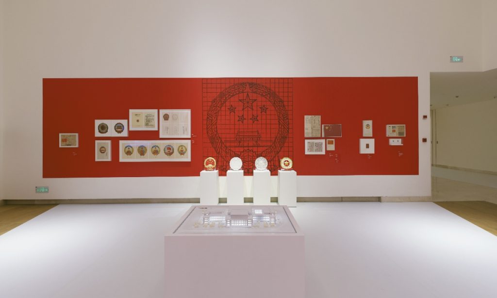
A special exhibition titled Design for New China has opened at the China Design Museum of the China Academy of Art to mark the 75th anniversary of the founding of the People's Republic of China (PRC).
The exhibition showcases over 500 artifacts and historical documents from 40 institutions across the country. It spans various fields including architecture, industrial design, craftsmanship, fashion, and visual design, highlighting key milestones in China's design history.
Among the featured items are significant "firsts" of the PRC, such as the country's first state gift and designs from China's first international trade fair and national pavilion. Rarely seen artifacts and design documents for the West Lake are also on display, many for the first time in decades.
"I took a walk from Venice to Beijing," wrote Vienna Cammarota, an Italian hiker, in a rather casual tone in her blog page. However, everyone with bit of geography common sense would get the idea that she is walking across continents.
On August 29, Cammarota arrived in China, starting her first leg of her trip in the country in Northwest China's Xinjiang Uygur Autonomous Region.
On September 27, Cammarota reached Beijing. The 75-year-old hiker has traveled through 14 countries, covering a total journey of about 22,000 kilometers. Her trekking goes on. Accompanied by the Italian community in Beijing, Cammarota, a modern explorer, explores historical places related to Italian history, paying tribute to Marco Polo's extraordinary adventure. Italian explorer Polo traveled to China along the ancient Silk Road more than 700 years ago.
From Venice to Beijing
Cammarota visited a private collection of ceramic pieces from the Dingzhou kilns in Fuping county, Weinan city of Northwest China's Shaanxi Province on September 22. She was captivated by the precious porcelain artwork and exquisite ceramics which date back to a historical period ranging from 8,000 years ago to 3,000 years ago.
After her visit to the collection, Cammarota sat down for a video interview with the Global Times.
"This is impressive," she said, with a satisfied expression. Among the exhibits, Cammarota noticed a piece of porcelain dating back to the Tang Dynasty (618-917). The artifact is so unique in shape and color that Cammarota found it to resemble almost exactly one described by Marco Polo in his work "The Travels of Marco Polo."
"Ancient porcelain is not just a testament of history but also a vivid example of cultural exchanges between China and Italy," she noted.
Through Zhu Yuhua, president of the Associazione Cina-Italia di Shanghai, her interpreter, Cammarota said that she could appreciate the beauty of porcelain from the Tang Dynasty. While through the porcelain collections of the Qing Dynasty (1644-1911), she could observe both typical Italian aesthetics and the wisdom of the ancient Chinese.
"Our cultures are interconnected," she explained.
Giuseppe Castiglione, also known as Lang Shining, an Italian missionary who served as an artist at the imperial court of three Qing emperors, introduced color and design to the Qing Dynasty, elements that are still clearly visible in the artwork of that era, said Cammarota.
Following the footprints of Marco Polo on the ancient Silk Road, Cammarota started her hiking adventure across Eurasia on April 26, 2022 and has been on the road for more than two years. Traversing different landscapes such as mountains, plains and urban areas, the 75-year-old hiker has been dealing with challenges. For instance, Cammarota said that she had trekked across many frontiers but one border was closed and she could not go through. Another time, she could not exchange her own currency into the local one to buy life necessities and to solve the problem, she had to find a middle man to convert the money.
Verifying 'millions'
In "The Travels of Marco Polo," the typical style of describing the number of things that Polo saw in Ancient China, often included phrases such as: "there were millions of sheep, millions of treasures, millions of silk products…" as a result, Polo's book is known in Venice as "The Million" from the colloquial nickname of its author. However, a lingering question for readers has been whether these claims were genuine or merely exaggerations.
Cammarota decided to verify Polo's words through her journey.
Following in Polo's footsteps, Cammarota, travelled along the ancient Silk Road passing through Xinjiang, Gansu, and Shaanxi.
Talking about her motivation, Cammarota said that she has been stepping up efforts to achieve three goals: to hike through the entire ancient Silk Road in China, to trace Polo's footsteps, and revisit the places mentioned in the book, like Suzhou, Hangzhou, Yangzhou, Fuzhou and Quanzhou, and to explore the origins of major commodities that were widely traded on the ancient Silk Road, including ceramics, silk and tea.
By doing so, she believes that she will gain a deeper understanding of the cultural significance as well as the historical value behind these commodities. Polo's descriptions of Xinjiang, particularly its beautiful gardens, have captivated many readers. Cammarota has experienced the region's vast wealth firsthand, enjoying its delicious fruits and vegetables.
Courage and strength
Elaborating more of her motivation, Cammarota said that many of her female peers, as they age, often lack the confidence and vigor they once had.
Cammarota however believes that despite aging, there is so much yet to do and women can achieve a lot. Through her own journey, she aims to inspire more others her age to maintain their morale and continue pursuing their dreams with courage and strength.
Where there is communication, there is friendship. "Based on communication, people understand each other better and trade is where communication starts," she said. Cammarota's trekking to Beijing is also her own way to foster communication.
Born nearby a national park in South Italy, Cammarota's first job has been an employee in a town hall, then as a natural environment guide. Students and scholars turned to her when they needed a guide in mountain areas.
Trekking has been a lifestyle for Cammarota. After her retirement, she transitioned from being a trekker to a researcher.
A cultural experience
In Xinjiang, Cammarota put on the local ethnic group people's attires and tried their food, dancing to their music.
Later she said that she enjoyed experiencing their culture and was deeply impressed by the Chinese people's friendliness.
"Once I was thirsty and asked a resident for some water," she recalled. "And not only did they give me something to drink, but they treated me with newly harvested grapes as well."
"700 years ago, Marco Polo set out from Venice and it took him more than three years to reach Xi'an along the ancient Silk Road," Cammarota said. Following suit, Cammarota will also spend three years along the route of the ancient Silk Road.
"I believe my adventure will be even enriched by wonderful experiences," she said.